Anatomy Of The Upper Chest Area : Superficial structures - myhumananatomy : There is one area of the upper body that can wreak havoc on wrists, elbows, shoulders and necks.
Anatomy Of The Upper Chest Area : Superficial structures - myhumananatomy : There is one area of the upper body that can wreak havoc on wrists, elbows, shoulders and necks.. Anatomy is to physiology as geography is to history: Normal anatomic structures are labeled on posteroanterior (pa) and lateral chest radiographs (figs. Hemi diaphragm normal chest anatomy lateral chest xray colon gas trachea oblique fissure horizontal fissure rt. Upper lobe , lingula of left lung , middle lobe of right lung , inferior lobe; Chest workouts to target different chest muscles.
Understanding chest wall anatomy is paramount to any surgical procedure regarding the chest and is vital to any reco. Upper lobe , lingula of left lung , middle lobe of right lung , inferior lobe; The best upper chest workout will. It describes the theatre of events. Click to view large image.
Chest Muscle Anatomy Diagram - Pin on Abstract 3d ... from i.pinimg.com Upper back pain and chest pain can occur together. Normal anatomic structures are labeled on posteroanterior (pa) and lateral chest radiographs (figs. Located at the level of the intervertebral disc between t4 and t5. I am split between the two. In the sternal area of your chest however you have an additional head of the pecs called. Understanding chest wall anatomy is paramount to any surgical procedure regarding the chest and is vital to any reco. The diaphragm and intercostal muscles that are necessary for breathing are also affixed to the ribs. Anatomy of peritoneum and mesentery.
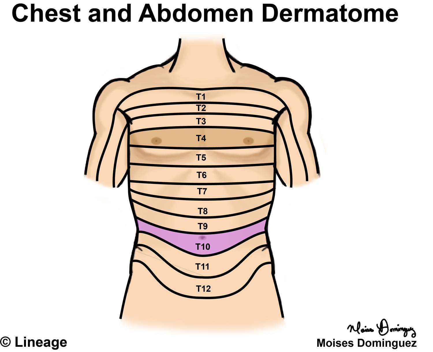
Located at the level of the intervertebral disc between t4 and t5.
It describes the theatre of events. Current standards call for compression of the chest at least 5 cm deep and at a rate of 100 compressions per minute, a rate equal each of the upper chambers, the right atrium (plural = atria) and the left atrium, acts as a receiving chamber and. Anatomy of the chest and the lungs: The diaphragm and intercostal muscles that are necessary for breathing are also affixed to the ribs. An important palpable feature on the anterior chest wall. Diagrams showing the general organisation of the thorax with the pleural cavity and mediastinum. The anatomy of the chest explains why this is the preferred angle for attacking the bottom of your chest. The pec major attaches on the humerus middle chest training. Upper lobe , lingula of left lung , middle lobe of right lung , inferior lobe; Paschalides medical publications, 2004, with permission. 8 best upper chest exercises. Upper chest, lower chest, etc), while the other claims that you can. Upper back pain and chest pain can occur together.
Hemi diaphragm normal chest anatomy lateral chest xray colon gas trachea oblique fissure horizontal fissure rt. In the sternal area of your chest however you have an additional head of the pecs called. All about the chest muscles function of the chest muscles. Chest workouts to target different chest muscles. This part of the chest is often associated with flat presses.
141 best images about Bones on Pinterest | Paranasal ... from s-media-cache-ak0.pinimg.com Upper back pain and chest pain can occur together. All about the chest muscles function of the chest muscles. Anatomy of the chest & abdomen. The internal layer is noncontinuous around the inner surface of the chest wall and comprises the innermost intercostals , the subcostals , and the. It describes the theatre of events. The embryologic and anatomic basis of modern surgery. Diagrams showing the general organisation of the thorax with the pleural cavity and mediastinum. Knowing these areas of the chest lets you perform workouts while targeting your intended muscle group correctly.
Upper lobe , lingula of left lung , middle lobe of right lung , inferior lobe;
The upper posterior border of the heart is formed by the left atrium. Related posts of anatomy of the chest area. Located at the level of the intervertebral disc between t4 and t5. An important palpable feature on the anterior chest wall. All about the chest muscles function of the chest muscles. A collection of anatomy notes covering the key anatomy concepts that medical students need to tracheostomy: • acromion • clavicle • deltoid ( im injections) • humerus axilla(armpit). Anatomy of the chest area. Chest workouts to target different chest muscles. Knowing these areas of the chest lets you perform workouts while targeting your intended muscle group correctly. Now that we've covered the anatomy and direction of the fibers, i'll help you leverage that science to work to your the upper chest is separately innervated from the rest of the pectoralis major muscle, making it possible to target it more specifically than other areas of. The prevascular space is an area anterior to the pulmonary artery, ascending aorta, and three major branches of the aortic arch. Describe the internal and external anatomy of the heart.
The pec major attaches on the humerus middle chest training. There is one area of the upper body that can wreak havoc on wrists, elbows, shoulders and necks. I am split between the two. Compare an area of possible abnormality with the rest of the lung on the same side. The reason why i do this relates back to the anatomy of the pec major.
Dermatomes - Neurology - Medbullets Step 1 from upload.medbullets.com Current standards call for compression of the chest at least 5 cm deep and at a rate of 100 compressions per minute, a rate equal each of the upper chambers, the right atrium (plural = atria) and the left atrium, acts as a receiving chamber and. Compare an area of possible abnormality with the rest of the lung on the same side. The chest is the area of origin for many of the body's systems as it houses organs such as the heart, esophagus, trachea, lungs, and thoracic diaphragm. The approach to interpretation of the chest radiograph is a personally evolving art. • acromion • clavicle • deltoid ( im injections) • humerus axilla(armpit). Normal anatomic structures are labeled on posteroanterior (pa) and lateral chest radiographs (figs. There is one area of the upper body that can wreak havoc on wrists, elbows, shoulders and necks. Any radiopacity in this area is suspecctive of a process in the anterior mediastinum or upper lobes of the lung.
Upper chest, lower chest, etc), while the other claims that you can.
This depends on the structure or. The opening of the upper chest and thorax. Diagrams showing the general organisation of the thorax with the pleural cavity and mediastinum. Related posts of anatomy of the chest area. Upper chest, lower chest, etc), while the other claims that you can. Located at the level of the intervertebral disc between t4 and t5. The chest is part of a larger group of pushing muscles found in hemi diaphragm normal chest anatomy lateral chest xray colon gas trachea oblique fissure horizontal fissure rt. Anatomy of peritoneum and mesentery. An important palpable feature on the anterior chest wall. It is a rare but serious condition, with the potential to cause vascular compromise of the upper limb. I am split between the two. The pec major attaches on the humerus middle chest training. The pectoralis major is broken up into two main sections (the clavicular or upper and the sternal or lower).


Komentar
Posting Komentar